Lesson 1
What is HER2?
Established science. Settled medical knowledge, curated for teaching — distinct from the platform’s own evolving research.
HER2 is a protein that sits on the outer surface of cells. Its full name is human epidermal growth factor receptor 2. Think of it as an antenna: it waits for a signal and, when it gets one, it tells the cell to grow and divide. In healthy tissue that’s a normal, useful job. The trouble starts when a cell has far too many of these antennas.
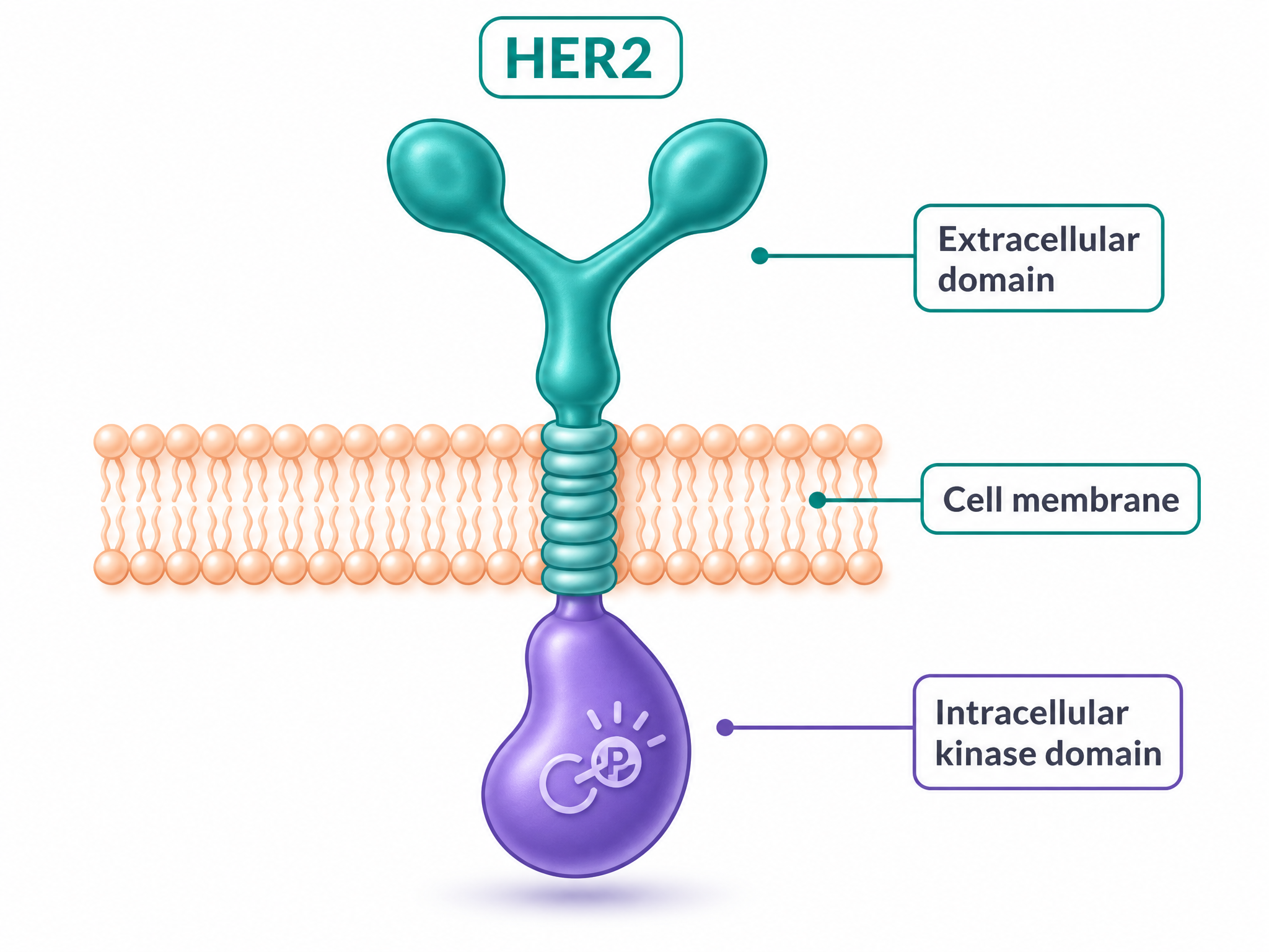
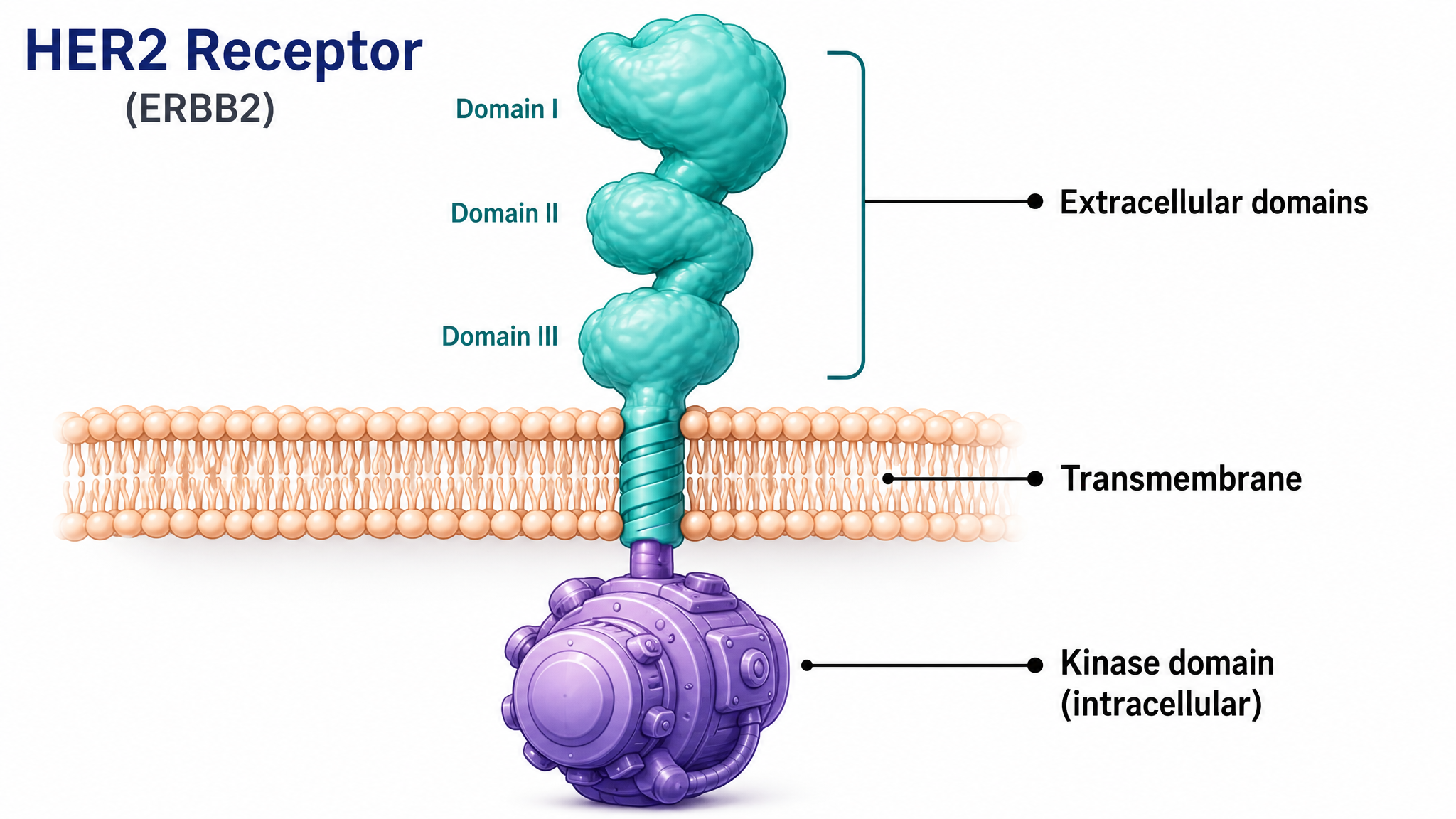
The part outside the cell is the receiver. The short piece crossing the membrane anchors it. The part inside the cell is the switch that fires the growth message onward. Hold on to those three parts — in the next lesson you’ll see that each treatment targets a different one.
The core problem
Too much of a normal thing
About one in five breast cancers — and some stomach and other cancers — are “HER2-positive.” That means the cancer cells make far more HER2 antennas than normal, sometimes a hundred times more. This is called overexpression.
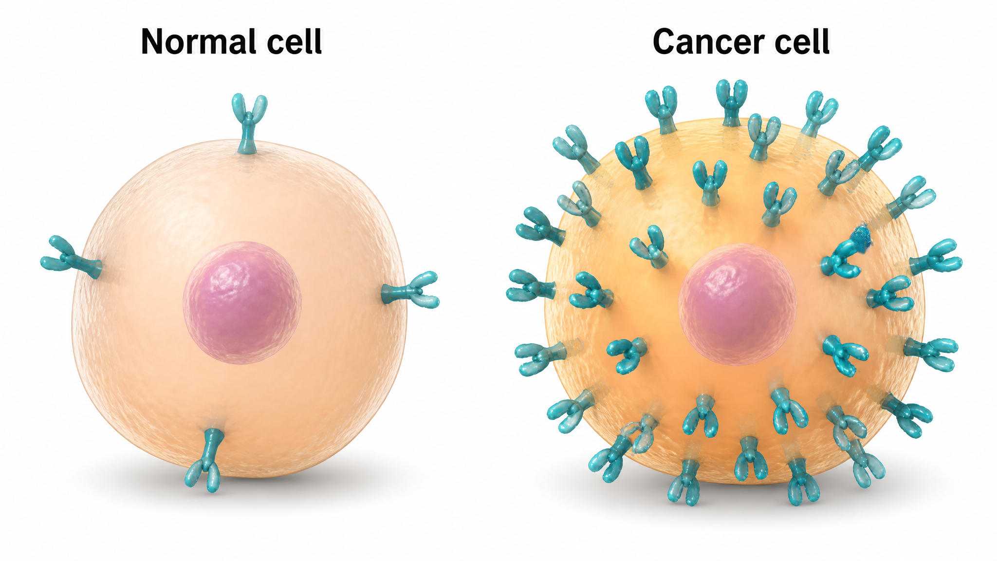
With so many antennas, the cell doesn’t need to wait for an outside signal anymore — the sheer crowding is enough to keep the growth message switched on. The result is a cancer that grows and spreads faster. The upside: because these cells lean so heavily on HER2, a drug that blocks HER2 hits the cancer hard while largely sparing normal cells.
HER2-positive
The cancer makes far too much HER2. These are the cancers HER2 medicines are designed for.
Why it matters
Because the cancer depends on HER2, switching HER2 off can stop the growth signal at its source.
How the signal travels
From the surface to the switch
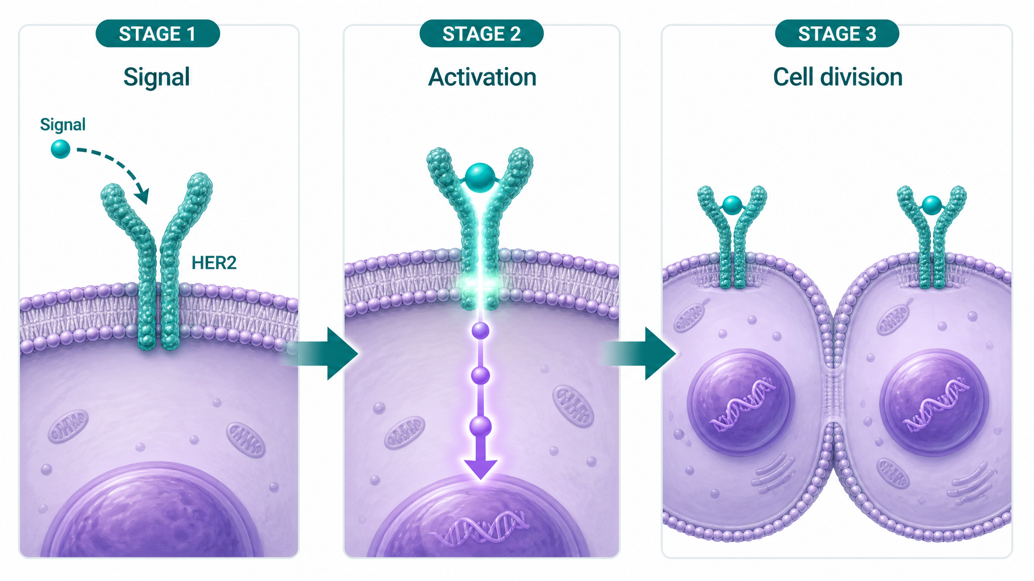
When HER2 is switched on, it sets off a relay inside the cell — one molecule flips the next, like a line of dominoes — ending in the command “grow and divide.” Scientists call these relays signaling pathways. You don’t need the names; the idea is what matters: one switch at the surface becomes a loud, repeated command deep inside the cell.
The key move
HER2 works in pairs
Here is the single most important detail for understanding the treatments. A HER2 antenna can’t fire on its own — it has to pair up with a neighbouring receptor first. Two receptors come together, and only then does the switch turn on. This pairing-up is called dimerization.
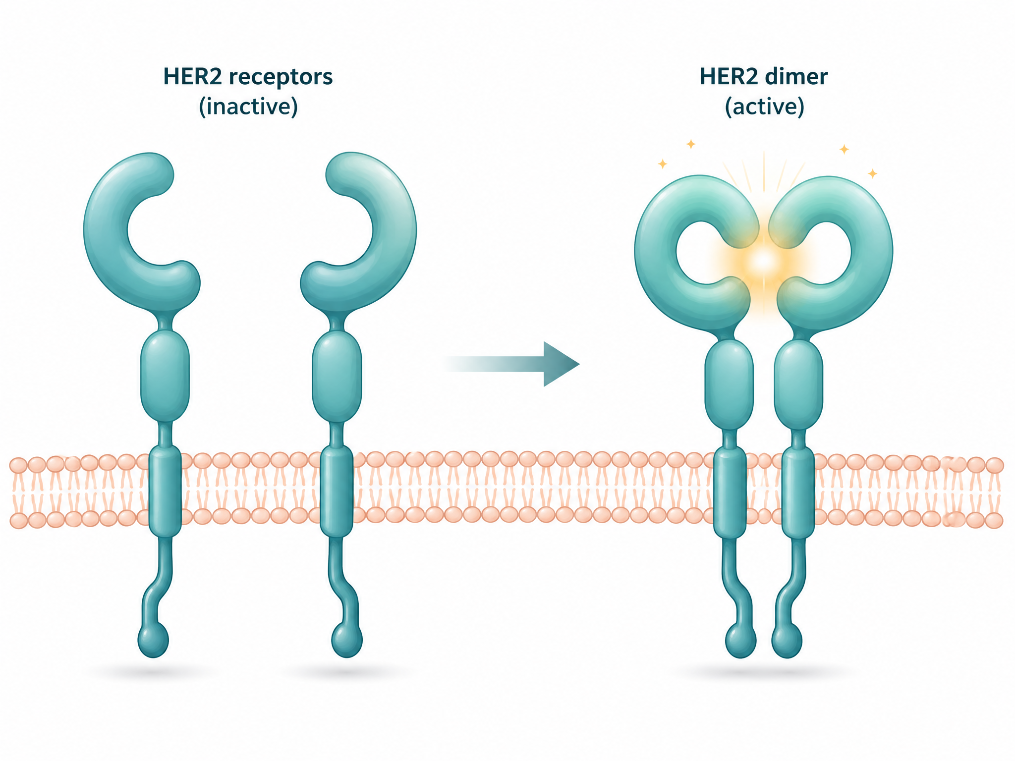
This is a gift for drug designers. If you can stop the receptors from pairing up, you stop the signal before it ever starts — and that is exactly what one of the major HER2 antibodies does. Keep dimerization in mind for the next lesson.
A map to remember
The parts, all in one picture
This labelled map pulls the whole receptor together. It’s the same diagram we’ll reuse in the next lesson to show where each treatment lands — so it’s worth a good look now.
The real thing
And here is the actual protein
Everything above is a simplified drawing. But HER2 is a real, physical molecule that scientists have measured atom by atom. Below is the genuine, experimentally-determined 3D structure of HER2’s outer region — the same data this platform studies, loaded live from the public protein databank. Give it a moment to appear; you can open the full explorer to turn it around yourself.
Real measured data · PDB 1N8Z
This is not a drawing. It is the outer region of HER2, solved by experiment and shared openly by researchers. The ribbon traces the actual folded chain of the protein. Seeing the real thing is a good reminder of what all the illustrations above are standing in for.
Beyond the settled science: the platform’s frontier
You now know what HER2 is and why too much of it drives cancer. That’s the settled part. What’s not settled — which mutations blunt which drugs, which new ideas hold up — is exactly what this platform tracks as it reads the latest research. That evolving layer is kept separate from this guide on purpose.